April 30, 2026
MoleMap Team

Skin cancer is the most common type of cancer in Australia and New Zealand – but it’s reassuring to know that, if found early, it can be successfully treated in the majority of cases.
Treatment for skin cancer will vary from person to person. It will depend on the type of skin cancer you’ve been diagnosed with, how big and deep the cancer is, and whether it has spread to other parts of your body. Some people receive one type of treatment, while others may need a combination of treatments.

Most skin cancers begin in your skin’s top layer, the epidermis. The epidermis contains three main types of cells – which is how the different skin cancers are classified.
Squamous cells: are in the outer part of the epidermis. The skin cancer that can form in these cells is called squamous cell carcinoma.
Basal cells lie beneath the squamous cells. They eventually move up the epidermis to become new squamous cells. Skin cancer that begins in basal cells is called basal cell carcinoma.
Melanocytes are cells found in the bottom layer of the epidermis. Skin cancer that begins in melanocytes is called melanoma.

As the diagram shows, the cells affected by each type of cancer lie in different layers under the skin. If left undetected, the cancer can potentially spread deeper into other organs or parts of the body.
Melanoma is less common than basal and squamous cell carcinoma – but it’s far more dangerous, because it can spread rapidly to other organs. It can become life-threatening within a few months if left to grow untreated. This is why early skin cancer detection and treatment is so vital.
Depending on your diagnosis, you may be treated by a number of different health professionals.
Your GP can sometimes perform simple surgery to remove moles and skin lesions. They can also provide you with a referral to a dermatologist, which is a doctor that specialises in diagnosing and treating skin diseases.
You may also be referred to a surgeon to remove your skin cancer. Depending on the stage of the cancer, or where it is on the body, this might be a general surgeon, a surgical oncologist, or a plastic surgeon (who is trained in reconstructive techniques).
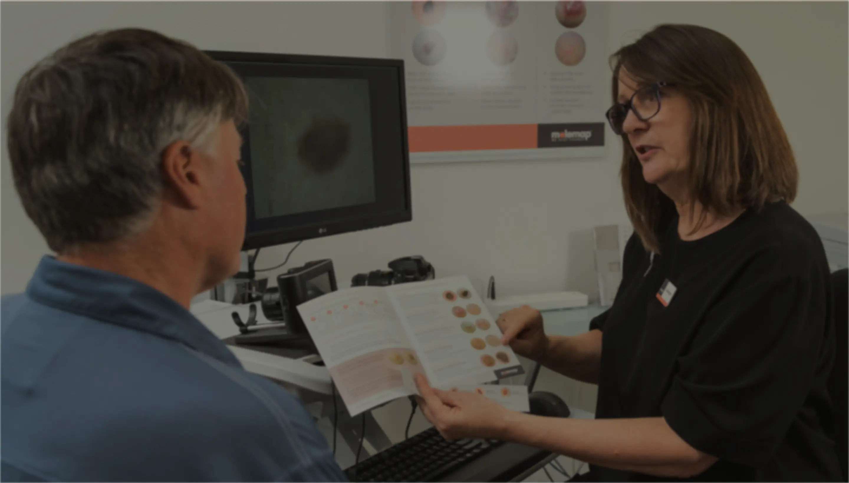
At MoleMap skin cancer clinic, your initial skin check is carried out by a trained melanographer. They undertake a thorough all-body check of your skin to detect any new or suspicious-looking moles or skin lesions. They can also advise you on the most effective preventive skin cancer treatment régime.
MoleMap also has team of dermatologists who review the photographic images taken by the melanographer, which means there are two set of expert eyes checking your skin.
If you have a suspicious-looking mole or skin lesion, prior to treatment, your GP or dermatologist may perform a biopsy. This is a quick procedure that only takes about 15 minutes. (Your doctor will inject a local anaesthetic to numb the area, so you won’t feel anything). They then use a biopsy punch, razor or scalpel to carefully remove a small sample of skin.
This sample is sent to a pathology lab, where it’s examined under a microscope by a pathologist (a specialist doctor who is trained to detect diseased cells). The pathology results are usually ready in about a week.
If the biopsy sample is found to be skin cancer, your dermatologist or GP will discuss the next treatment steps. The treatment you undergo will depend on the type of skin cancer you have, and what stage the cancer is at.
In the past, doctors would often surgically remove moles as a precaution – even the completely benign ones. This meant patients were sometimes undergoing a procedure (and getting scars) unnecessarily. On the other hand, dangerous moles were often missed during a visual-only check and left untreated.
MoleMap was launched by a group of doctors in 1997 who wanted to provide a more targeted and accurate assessment of potential skin cancers.
The most common treatment for skin cancer is to remove the cancer, usually under a local anaesthetic. This procedure may be performed by your GP, by a dermatologist or you may be referred to a surgeon (such as a general surgeon or plastic surgeon).
Using a scalpel, the Dermatologist or surgeon removes the mole or skin lesion, along with a “safety margin” of surrounding skin. This is sent to a pathology lab for analysis. The affected area is usually stitched together and may result in a small scar.
Often the entire skin cancer is removed from the initial procedure, and no further treatment is needed. However, if the lab finds cancer cells beyond the margins, surgery may be performed again, until the margins are found to be cancer-free.
With Melanoma often the doctor will remove first time with relatively small margins and once the pathologist has determined the thickness of the skin cancer, the doctor will undertake a wide local excision to obtain the best safety margins for the patient.
Mohs surgery is a specialist procedure where the cancer is removed ‘layer by layer’ in a single visit. The surgeon removes a layer of tissue, immediately examines it under a microscope, and then removes another layer if necessary. The advantage of this technique is that it preserves as much healthy tissue as possible, and can minimise scarring.

In addition to cutting out the cancerous mole or lesion, there are a number of other treatment options.
Cryotherapy (or cryosurgery) is used by dermatologists to treat a range of skin problems, including some cancers. With this procedure, liquid nitrogen is sprayed on to the area of skin to freeze it. This light freezing causes the skin to blister, scab and fall off. Cryotherapy is more often used to treat pre-cancerous lesions such as solar keratosis, a pre-cancerous lesion. Curettage (scraping), cautery (burning) and chemical peeling are other techniques which have a similar ‘blistering over’ effect.
Certain topical ointments and creams are also used to treat superficial skin cancers. Generally these are prescribed chemotherapy topical medicines that can be directly applied to the skin, instead of being given by mouth or injected into a vein. It’s usually applied once or twice a day for several weeks.
Photodynamic therapy uses a combination of a red-light laser and a cream. In Australia, it is an approved treatment for solar keratoses and basal cell carcinoma, and success rates are around 80 – 85%.
Radiation therapy is sometimes used when the area of skin is difficult to treat with surgery – such as around the eye, eyelid, ear or tip of the nose.
All types of skin cancer have the potential to spread (or ‘metastasise’) to other parts of the body but it’s not very common. Melanoma, if caught early and it’s thin, is unlikely to spread, but if left untreated it can get deeper into the skin and thus have a potential to spread via blood vessels or lymphatic system. Squamous cell carcinomas can also have a potential to spread but that risk is very low.
If caught early, 90% of melanomas can be cured with simple surgery alone. This is the most common treatment for melanomas.
For skin cancers with a high risk of spreading or metastasising, surgery may be followed by other treatments.
If you have been diagnosed with advanced melanoma, or another type of skin cancer which has spread, you may undergo a combination of different treatments. This will often include surgery, drug therapy (such as chemotherapy or immunotherapy), and radiation.
When detected early, skin cancers are relatively easy to treat and have an excellent prognosis. Our mole check service includes precision mole monitoring to detect even the smallest changes in your skin over time.
References:
1. Cleveland Clinic: Skin Cancer https://my.clevelandclinic.org/health/diseases/15818-skin-cancer#:~:text=Nearly%20all%20skin%20cancers%20can,for%20a%20professional%20skin%20checkup;
2. NPS Medicine Wise: Non-surgical treatment for melanoma, https://melanoma.org.au/for-patients/melanoma-treatment;
3. Melanoma Institute Australia: https://melanoma.org.au/for-patients/melanoma-treatment/; 4. Skin Cancer Foundation: Melanoma Treatment https://www.skincancer.org/skin-cancer-information/melanoma/melanoma-treatments/